
Publications
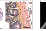
Our first work on the enteric nervous system, the “brain in the gut” is out!
Our first work on the enteric nervous system, the “brain in the gut” is out! Check out out work at bioRxiv: https://www.biorxiv.org/content/10.1101/2022.06.15.496266 UPDATE (2023/01) : The article has been accepted in Frontiers in Neuroanatomy ...
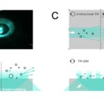
Understanding the effect of scattered evanescent light
Evanescent-wave scattering is a topic in classical electrodynamics and in the study of colloidal particles near a boundary. However, how such near-surface scattering at sub-cellular refractive-index heterogeneities degrades the excitation confinement ...
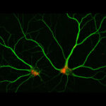
A nutrient sensor in the cell’s stomach | INSB (cnrs.fr)
© Solène Lebrun & Bruno Gasnier To stay in good health, cells that no longer divide such as neurons renew their components by degrading them and recycling their building blocks in acidic compartments called lysosomes. These “micro-stomachs” ...
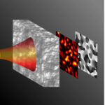
Complex scattered light-field measurement with a simple wavefront-sensor
Wavefront sensors are simple and robust phase sensors at the basis of adaptive optics in astronomy and bio-imaging. Adaptive optics has been introduced in bio-imaging to compensate smooth aberrations mostly arising from the refractive index mismatch a ...
A simple, inexpensive and multi-scale 3-D fluorescent test sample for optical sectioning microscopies
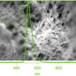
Pyranine-labeled tissue paper, imaged upon 405-nm epifluorescence excitation through a 455LP LP dichroic and 465LP emission filter. Objective ×20/NA0.25. Overlaid are the normalised absorbance (dashed) and emission spectra (through line), respectively. I ...
Heating, not highly non-linear photo damage is limiting for 920-nm 2-imaging
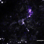
Unraveling how neural networks process and represent sensory information and how these cellular signals instruct behavioral output is a main goal in neuroscience. Two-photon activation of optogenetic actuators and calcium (Ca 2+) imaging with gen ...
Spectral correlations in forward scattering media
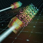
Artist illustration of chromatic speckle expansion after a scattering sample. (© Clément Molinier) Imaging deep in scattering tissues has been typically achieved by exciting non-linear optical processes thanks to broadband ultrashort laser pulses. Scatte ...
Evanescence in emission: seeing fluorescence from supercritical angles
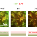
Read it here : Supercritical angle microscopy and spectroscopy Authors: Martin Oheim, Adi Salomon, Maia Brunstein Abstract: Fluorescence detection, either involving propagating or near-field emission, is widely being used in spectroscopy, sensing and micro ...
How to calibrate evanescent-wave penetration depths for biological TIRF microscopy?
Total internal reflection sets up an evanescent wave that – when used for microscopic imaging – renders the microscope short-sighted and enables the selective observation of the near-membrane space. But how deep is deep? In this Biophysical Pe ...
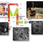
Fast volumetric Imaging of Brain Organoids With a New Single-Objective Planar-Illumination Two-Photon Microscope
Imaging large tissue volumes at high spatio-temporel resolution has been a major challenge for scanning-type 2-photon microscopes. Now, SPPIN researchers around Martin Oheim and from TILL.id (Munich) designed and successfully validated a new microscope sch ...