
News, Publications
Fast volumetric Imaging of Brain Organoids With a New Single-Objective Planar-Illumination Two-Photon Microscope
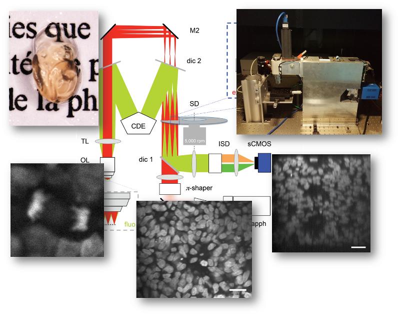 Imaging large tissue volumes at high spatio-temporel resolution has been a major challenge for scanning-type 2-photon microscopes. Now, SPPIN researchers around Martin Oheim and from TILL.id (Munich) designed and successfully validated a new microscope scheme that combines features from both confocal laser-scanning and light-sheet microscopes: a micrometric optical sectioning capacity and sub-micrometric spatial resolution, a large field of view and high frame rate, and a low degree of invasiveness. Their EU-fiananced work (H2020 Eureka! EUROSTARS “OASIS”), on uncleared and cleared 3-D cultures of hIPSCs as a sample, is now published in Frontiers in Neuroanatomy (Rakotoson*, Delhomme*, Djian* et al. (2019) Front. Neuroanat. 13:77. doi: 10.3389/fnana.2019.00077)