Transverse research lines of the SPPIN are collaborative research efforts in which at least three labs of the SPPIN team up to provide a shared research infrastructure that could not easily be bought or maintained by a single lab. At present, these efforts include, i- the M-Cube light-sheet mesoscope, the preparation of hIPSC organoids and the development of a universal, extremely rapid and non-noxious tissue clearing technique. This website gives a work-in-progress view of developments combining these three axes, under the umbrella term “Multiscale neuroanatomy”. Stay tuned.
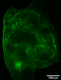
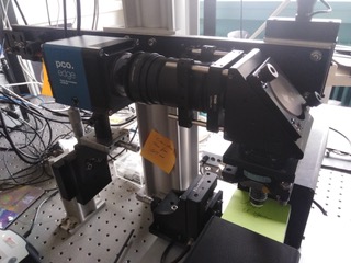
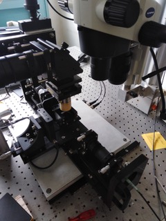
Tissue clearing techniques, along with progress in 3-D fluorescence imaging have prompted a revival of neuroanatomy. The workhorse in such studies has been the light-sheet microscope, the defining feature of which is a crossed arrangement of excitation and collection optics. This arrangement brings about a trade-off between field-of-view against spatial resolution. Brain structures, however, span spatial scales of more than 6 orders of magnitude — from blood vessels and neuronal networks linking areas centimeters apart to tiny compartments like dendritic spine shafts or astrocyte peripheral processes that measure little more than a few tens of nm. Imaging such diverse structures in a continuous manner, from overviews to sub-µm detail would be extremely useful, but it would require a continuous adaptation in the illumination and collection optics that is difficult to implement on a single instrument. We built a compact, modular light-sheet microscope designed for correlative micro- meso- and macroscopic observation (M-Cube).